Submitted by jenna on Mon, 06/24/2013 - 23:53
Hi experts,
I am running a resting state FC analysis, and am confused at my FC maps resulting from REST. I have attached an image. It looks as though the FC map is not including many inferior regions of the brain. I was wondering....does this image look normal, or could it be that something went wrong. I used a mask created with TOM to mask my activation....but I resliced this mask according to my functional images....so they are in the same size. Could this be due to a problem with the voxel sizes, or is it just a bug in the program? I am skeptical to continue from here for I fear the analysis will not include the inferior regions, which I will be focusing on.
Thanks,
Jenna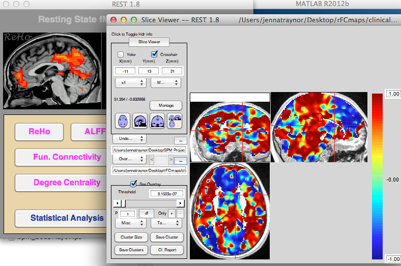
Submitted by ZangYF on Sat, 06/29/2013 - 06:06 Permalink
Re: FC map problems
Please check the size of "bouding box" you selected. Did you use SPM for data prerocessing or DPARSF?
Submitted by jenna on Sat, 06/29/2013 - 10:30 Permalink
Re: FC map problems
Hi, thanks for your response. I used SPM to preprocess and the size of the bounding box was -78 to 78, -112 to 76 and -50 to 85. This was the default setting. Does the REST program require specific measurments? Thankyou!
Submitted by YAN Chao-Gan on Mon, 07/01/2013 - 23:09 Permalink
Re: FC map problems
Hi,
1. The default bounding box of SPM is smaller. You can check the default one in DPARSF.
2. Could you check your mask first with REST Slice Viewer AND MRIcroN first?
Best,
Chao-Gan
Submitted by jenna on Wed, 07/03/2013 - 20:54 Permalink
Re: FC map problems
Hi Chao-Gan,
Thanks for your response. When I set my bounding box to DPARSF default values, my FC map generates activation completely outside of the brain. And even when I run my analysis with no mask, the result is the same. I am going to send my images to the other person who has offerred to help and hopefully we can figure out what is going on.
Thankyou so much!
Jenna
Submitted by ZHANG_RESTadmin on Thu, 07/04/2013 - 16:44 Permalink
Re: FC map problems
Hi Jenna,
from the images i thought its possible that your coreg and segmentation had problems.
your raw fMRI data looks fine, but after preproc, the image is not in the standard space (MNI space).
Please use SPM checkReg to see if your functional image is perfectly coreg to your T1 image, and see if your segmntation was good (your gm, wm images should be all registered to spm apriori gm, wm templates).
Han
Submitted by YAN Chao-Gan on Tue, 07/09/2013 - 04:09 Permalink
Re: FC map problems
And if not, checking "Reorient Fun*" and "Reorient T1*" checkboxes may help.
Best,
Chao-Gan
Submitted by ZHANG_RESTadmin on Wed, 07/03/2013 - 17:01 Permalink
Re: FC map problems
Hi Jenna, I am very interested why you got this FC map. It seems your bottom brain FC totally lost.
Can you send me one of your original raw functional image? And can you show your mask file as well?
Usually in SPM, mask file will be generated in task activation detection. But from your description I was confused about your FC map generation and mask file generation. Can you be more specific?
To send the image file, please refer to my email: napoleon1982@gmail.com
I will post the reply here to tell you and others what happened if I find the answer.
Han
Submitted by jenna on Wed, 07/03/2013 - 20:55 Permalink
Re: FC map problems
Hi Han,
Thanks for your response. I will send you that information, and more information about my mask, sometime today. I did not create the mask in SPM, I used a toolbox called template-o-matic to mask my results. Maybe I will also send you the mask.
Thankyou so much for your help!
Jenna