Region definition: The region lateral to the posterior convolutions of the insula for its internal part and by the union of the precentral and postcentral gyri after the Rolandic sulcus ends for its lateral part. References Dejerine (1980).
Jason W. Bohland mail, Hemant Bokil, Cara B. Allen, Partha P. Mitra
Abstract
Many neuroscientific reports reference discrete macro-anatomical regions of the brain which were delineated according to a brain atlas or parcellation protocol. Currently, however, no widely accepted standards exist for partitioning the cortex and subcortical structures, or for assigning labels to the resulting regions, and many procedures are being actively used. Previous attempts to reconcile neuroanatomical nomenclatures have been largely qualitative, focusing on the development of thesauri or simple semantic mappings between terms. Here we take a fundamentally different approach, discounting the names of regions and instead comparing their definitions as spatial entities in an effort to provide more precise quantitative mappings between anatomical entities as defined by different atlases. We develop an analytical framework for studying this brain atlas concordance problem, and apply these methods in a comparison of eight diverse labeling methods used by the neuroimaging community. These analyses result in conditional probabilities that enable mapping between regions across atlases, which also form the input to graph-based methods for extracting higher-order relationships between sets of regions and to procedures for assessing the global similarity between different parcellations of the same brain. At a global scale, the overall results demonstrate a considerable lack of concordance between available parcellation schemes, falling within chance levels for some atlas pairs. At a finer level, this study reveals spatial relationships between sets of defined regions that are not obviously apparent; these are of high potential interest to researchers faced with the challenge of comparing results that were based on these different anatomical models, particularly when coordinate-based data are not available. The complexity of the spatial overlap patterns revealed points to problems for attempts to reconcile anatomical parcellations and nomenclatures using strictly qualitative and/or categorical methods. Detailed results from this study are made available via an interactive web site at http://obart.info.
Submitted by ZangYF on Thu, 02/13/2014 - 17:17 Permalink
Re: matlab和rest报告的峰值点BA脑区不一致
insula在原来的Brodmann Area是没有编号的。在文章中,最好加上采用具体的什么方法报告的脑区名称,因为不同软件对脑区定义并不是完全。但我对具体的操作不清楚。
Submitted by ZHANG_RESTadmin on Tue, 02/18/2014 - 14:28 Permalink
Re: matlab和rest报告的峰值点BA脑区不一致
建议报出insula的哪个部位。而不是只报BA区。两个软件结果均不可尽信。
Submitted by water on Wed, 02/19/2014 - 16:44 Permalink
Re: matlab和rest报告的峰值点BA脑区不一致
谢谢张老师,发现这两个软件对同一个峰值点BA分区不一致的时候挺多。
Submitted by water on Sat, 02/22/2014 - 09:39 Permalink
Re: matlab和rest报告的峰值点BA脑区不一致
谢谢张老师,今天仔细看了下rest的BA模板,其中没有13区,所以rest不可能显示13区
Submitted by admin on Thu, 02/20/2014 - 00:35 Permalink
Re: matlab和rest报告的峰值点BA脑区不一致
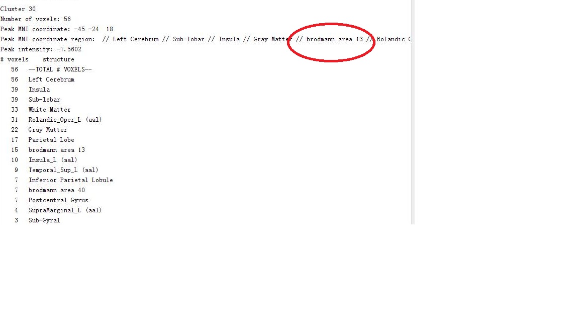
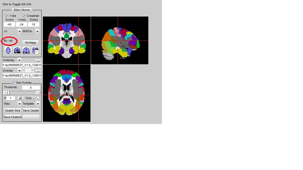
你的结果1显示的是一个cluster的结果,其中BA13有15个voxels,BA13座标应该不是你的peak座标。
结果2中是peak 点对应的分区 Rolandic_Oper_L。
我没有看到有什么不一致
Submitted by water on Thu, 02/20/2014 - 17:18 Permalink
Re: matlab和rest报告的峰值点BA脑区不一致
宋老师,peak MNI coordinate region 这一行不是峰值点坐标所在脑区吗?从左到右脑区依次精确?
Submitted by admin on Fri, 02/21/2014 - 11:20 Permalink
Re: matlab和rest报告的峰值点BA脑区不一致
是啊,我也是这么理解的,peak应该是在最右Rolandic_Oper_L。
我不清楚这个cluster report生成的方法,看起来好像AAL和Brodmann间没有重合,实际上应该有不少重合。
Reference:
http://qnl.bu.edu/obart/region/AAL/17/CB/CYTO/HO/ICBM/LPBA/TALc/TALg/TG/
Rolandic_Oper_L (AAL)
Hemisphere: Left
Region definition: The region lateral to the posterior convolutions of the insula for its internal part and by the union of the precentral and postcentral gyri after the Rolandic sulcus ends for its lateral part. References Dejerine (1980).
Volume as defined: 7939 mm3
Standardized name: rolandic operculum
Submitted by water on Fri, 02/21/2014 - 20:45 Permalink
Re: matlab和rest报告的峰值点BA脑区不一致
谢谢宋老师,学习一下这个网站,无论用哪个软件的结果都要标记清楚
Submitted by admin on Fri, 02/21/2014 - 11:32 Permalink
Re: matlab和rest报告的峰值点BA脑区不一致
The Brain Atlas Concordance Problem: Quantitative Comparison of Anatomical Parcellations
Jason W. Bohland mail, Hemant Bokil, Cara B. Allen, Partha P. Mitra
Abstract
Many neuroscientific reports reference discrete macro-anatomical regions of the brain which were delineated according to a brain atlas or parcellation protocol. Currently, however, no widely accepted standards exist for partitioning the cortex and subcortical structures, or for assigning labels to the resulting regions, and many procedures are being actively used. Previous attempts to reconcile neuroanatomical nomenclatures have been largely qualitative, focusing on the development of thesauri or simple semantic mappings between terms. Here we take a fundamentally different approach, discounting the names of regions and instead comparing their definitions as spatial entities in an effort to provide more precise quantitative mappings between anatomical entities as defined by different atlases. We develop an analytical framework for studying this brain atlas concordance problem, and apply these methods in a comparison of eight diverse labeling methods used by the neuroimaging community. These analyses result in conditional probabilities that enable mapping between regions across atlases, which also form the input to graph-based methods for extracting higher-order relationships between sets of regions and to procedures for assessing the global similarity between different parcellations of the same brain. At a global scale, the overall results demonstrate a considerable lack of concordance between available parcellation schemes, falling within chance levels for some atlas pairs. At a finer level, this study reveals spatial relationships between sets of defined regions that are not obviously apparent; these are of high potential interest to researchers faced with the challenge of comparing results that were based on these different anatomical models, particularly when coordinate-based data are not available. The complexity of the spatial overlap patterns revealed points to problems for attempts to reconcile anatomical parcellations and nomenclatures using strictly qualitative and/or categorical methods. Detailed results from this study are made available via an interactive web site at http://obart.info.