-
Fri, 04/12/2019 - 22:44jiaxizethere are a large number of robots registered in BBS,therefore, we add the function of slider verification code. We are very sorry for that. Briefly, In the pop-up window, hold down the slider below and drag to complete the puzzle the detailed explanation is...16332 reads
-
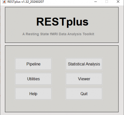 Sun, 01/24/2016 - 17:42REST-GroupRESTplus 从 REST(静息状态 fMRI 数据分析工具包)演变而来。它基于 Matlab 和 SPM8。RESTplus 包括四个主要模块,即管道、统计分析、实用程序和查看器。 The pipeline (either fixed or flexible) module provides a very easy way for data processing. After arrangethe DICOM or NIFTI filesand click a...76173 reads
 -
Sat, 05/25/2019 - 16:20jiaxize为了确保数据处理的可信性,我们拟寻找数据重复合作者。合作大致协议如下: 1、 我们将把我们的原始数据和数据处理的每一个过程发给您,由您独立完成数据处理的每个过程,然后与我们的结果进行比对,确保结果相同。 2、 数据重复合作者列在作者中(如果有导师指导,可以列2人)。 3、 数据重复合作者无权将这些原始数据用于其它任何用途。 4、 数据重复合作者需要具备与相应的研究比较相关的数据处理能力。 5、 需要在协商的时间内完成数据处理。 6、 双方协商之后签署合作协议。 本次需要重复的,...13642 reads
-
Tue, 04/08/2014 - 08:25Wei LiaoDynamic brain connectome (DynamicBC) analysis toolbox is a Matlab toolbox to calculate Dynamic Functional Connectivity (d-FC) and Dynamic Effective Connectivity (d-EC). Sliding window analysis (Bivariate Pearson correlation and Granger causality) and time...159067 reads
-
Wed, 01/18/2023 - 00:11JiaweiSunDear all, We are looking for another new Postdoc to work in an interdisciplinary and internationally competitive environment at Karolinska Institute, one of the top medical universities in the world and the home for the Nobel Prize in Medicine. We combine...10344 reads
-
Wed, 10/02/2019 - 14:50fanfan第二十二届全国心理学学术大会多模态神经影像专题工作坊招生简章(10月15日-10月18日,4天) 主办单位:中国心理学会 杭州师范大学心理科学研究院 承办单位:杭州拾睿科技有限公司 重要更新:根据大家的建议,我们在原来的2天讲座基础上,增加了为期2天的实操上机学习班,内容涵盖“state”任务和静息实验设计及相关数据分析实操入门、任务态fMRI数据处理实操入门以及基于坐标或体素的Meta分析介绍,价格仍然维持1500元。 一、工作坊简介: 近年来,磁共振影像技术的发展日新月异,...15162 reads
-
Sat, 08/17/2019 - 13:20jiaxize在当代的心理学研究中脑实验程序的编制已成为大多数的研究不可或缺的步骤。尽管在业界同仁的努力下开发出了基于MATLAB(或免费的Octave)的Psychtoolbox这样免费且功能强大的实验编程平台。但该平台的使用需要用户有较为丰富的编程经验,因而给使用者带来了巨大的使用成本。很多心理学研究工作者由于缺乏编程基础,往往会在编写实验程序中或者在学习编写实验程序上浪费大量宝贵的时间或者转而选择一些价格昂贵且功能单一的商业软件。 为了解决这一困扰很多心理学科研工作者的问题和促进实验编程的标准和简易化,...11475 reads
-
Wed, 09/05/2012 - 12:15REST-GroupResting-State fMRI Data Analysis Toolkit (REST) is a convenient toolkit to calculate Functional Connectivity (FC), Regional Homogeneity (ReHo), Amplitude of Low-Frequency Fluctuation (ALFF), Fractional ALFF (fALFF), Gragner causality, degree centrality,...248363 reads
-
Mon, 03/18/2019 - 00:02Icey各位老师好,有几个问题想请教: 1.使用dpabi做VBM,是否就按照dpabi里的VBM模块来处理就可以了呢? 2. 产生的smwc1*.nii是对GM images 进行modulated生成灰质的绝对体积吗?那如何才能得到用个体的体积校正后的值呢? 3. 关于reorient,它是一个optional的选项,所以是不是有可能只有某个被试的图像需要reorient,而不是整个群体一起做?那有没有什么具体的指标和方法来确定需不需要做reorient呢? 谢谢老师, Icey17360 reads
-
Tue, 03/12/2019 - 11:04asce老师您好,对于gretna这个工具箱,学生想请教您几个问题: 1. gretna 是否只能去除头动过大的体积,而不去除被试; 2. 对于网络分析部分的输入矩阵是得到的功能连接矩阵吗,还是Fisher‘z变化后的相关矩阵; 3.对于Modular Interaction这一网络属性中Community Index选项的输入是nodal index这个1-90的一列数吗? 望老师能不吝赐教!21915 reads